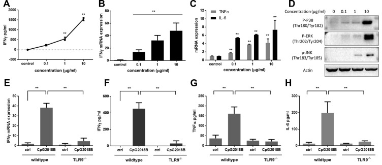
Figure 2.
CpG2018B stimulates IFN-γ and other cytokine secretion via TLR9. After incubation with different concentrations of CpG2018B for 48 hours, (A) IFN-γ was detected. (B and C) IFN-γ, TNFα and IL-6 mRNA levels were detected separately. (D) The phosphorylation of P38, ERK and JNK was detected by Western blotting. (E and F) In TLR9 knockout mice, pDCs were isolated and incubated with CpG2018B for 48 hours. IFN-γ secretion and mRNA levels were detected separately. (G and H) TNFα and IL-6 in pDCs were evaluated by ELISA. **p<0.01.

