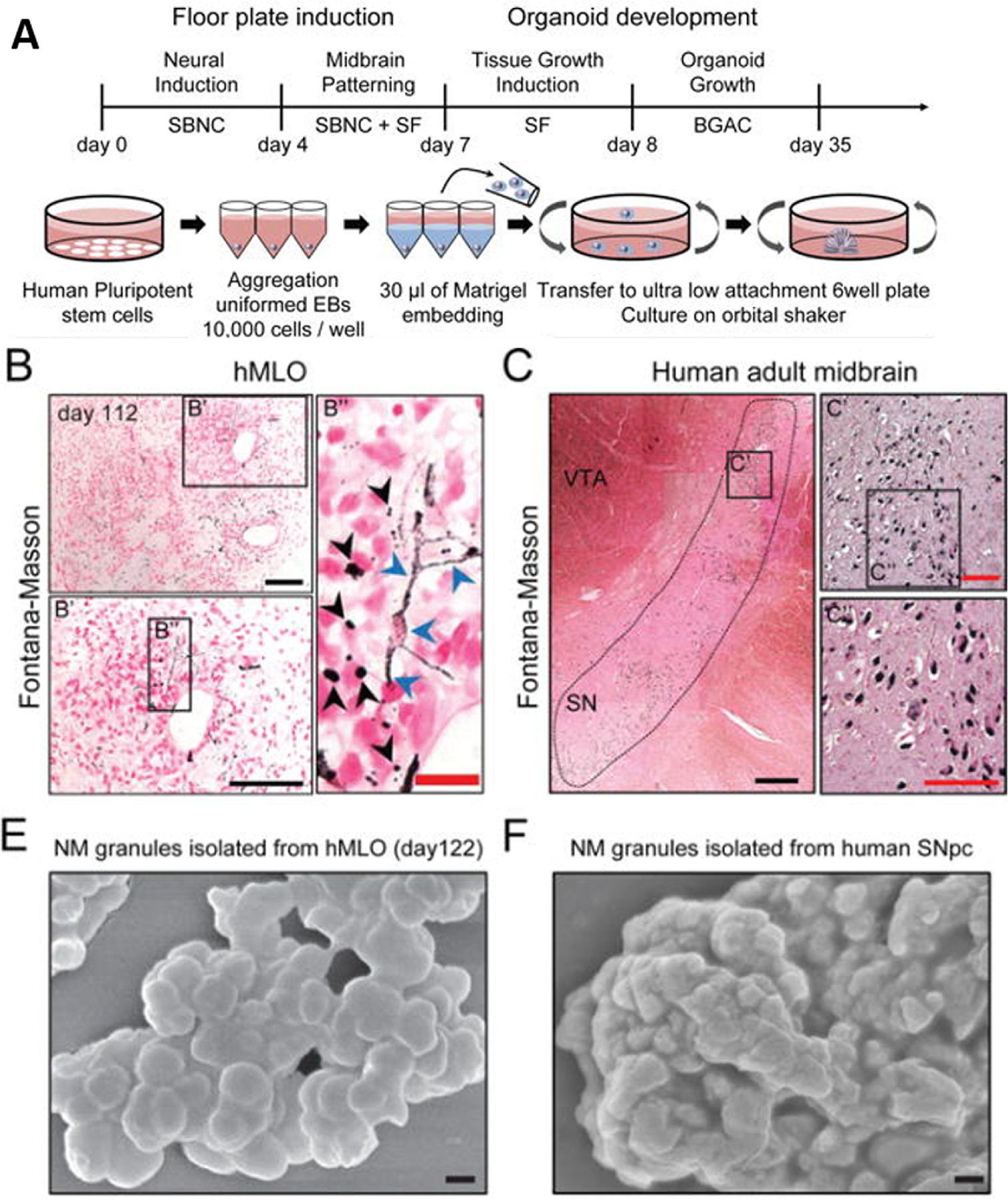
Figure 17. Midbrain Organoid.

(A) Schematic diagrams illustrating the overall strategy to midbrain organoids.
(B) Fontana-Masson staining to reveal NM-like granules in both intra- (blue arrows) and extracellular compartments (black arrows) of the organoids compared with (C) Fontana-human midbrain tissue. (E) SEM image of isolated NM granules from the organoids, compared with (F) Huma midbrain tissue. (Reprinted with permission from ref244. Copyright 2016 Cell Stem Cell).
