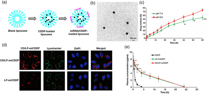
FIGURE 2.

Overview of experimental pathway for CDDP and miRNA loading. (a) The CDDP was loaded in the liposome shell while miRNA on the surface. (b) Morphology detection of CD/LP‐miCDDP with transmission electron microscopy. (c) Drug release study of CD/LP‐miCDDP in Ph 7.4 and Ph 5.0 conditions. (d) Cellular uptake analysis of CD/LP‐miCDDP and LP‐miCDDP in cervical cancer cells. (e) Plasma concentration–time analysis of CDDP, LP‐miCDDP and CD/LP‐miCDDP. *p <0.05, **p <0.01, ***p <0.0001. Copyright © 2020 SpringerOpen
