
FIGURE 5.
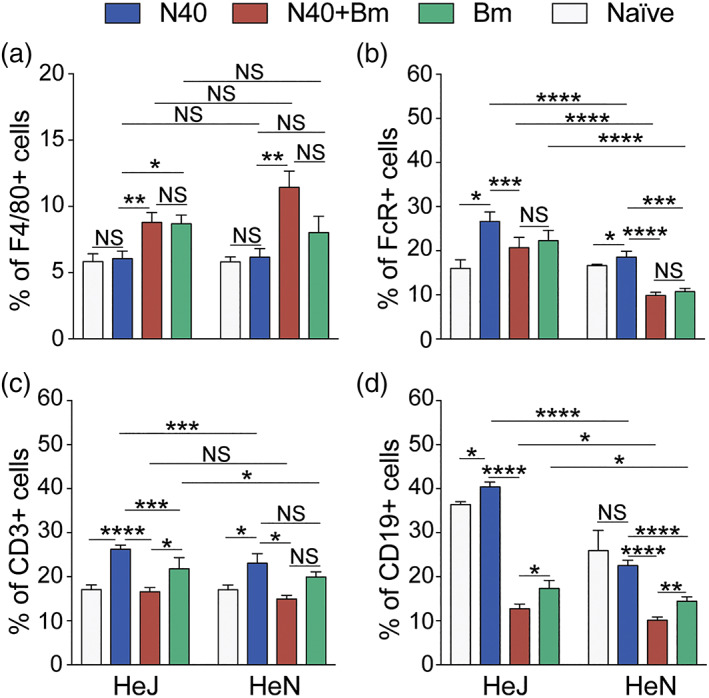
Modulation of splenic immune responses in mice by infection with B microti and N40 separately, or simultaneously. (a) Single cell suspension of mice spleen infected with N40, Bm and N40 + Bm were prepared and stained with fluorophore‐conjugated antibodies against F4/80+ (macrophages), (b) FcR+ phagocytic cells, (c) CD3+ (T cells), and (d) CD19+ (B cells), followed by FACS analysis. Higher percentages of B, FcR+ phagocytic and T cells were observed in C3H/HeJ mice. Proportions of various immune cells from spleens of mice from each experimental group analysed by FACS are expressed as mean ± SD. Statistical analysis was conducted using a two‐tailed unpaired student t tests for unequal variance to determine significant difference between the paired groups (NS‐Not Significant, *p < .05, **p < .01, ***p < .001, ****p < .0001)