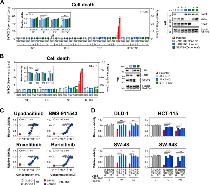
Fig. 3. JAK1/2 inhibition protects IECs from IFN-γ/TNF-α.
A Parental, JAK1, JAK2 or STAT1 knockout HT-29 cells were treated with IFN-γ and/or TNF-α (10 ng/ml each) for up to 72 h. SYTOX Green/Annexin V signals were recorded at indicated times, and relative viability was measured at 72 h (inner graph). Two knockout clones per each target (labelled as 'a' and 'b') were generated. Western blot validation of target knockout (right). B Parental, JAK1, JAK2 or STAT1 knockout DLD-1 cells were treated with IFN-γ and/or TNF-α (100 ng/ml each) for up to 72 h. SYTOX Green/Annexin V signals were recorded at indicated times, and relative viability was measured at 72 h (inner graph). Western blot validation of target knockout (right). C Relative viability of HT-29 cells pretreated for 1 h with vehicle (DMSO), JAK1-selective compound (upadacitinib), JAK2-selective compound (BMS-911543) or dual JAK1/2 inhibitors (ruxolitinib or baricitinib) starting at 10 μM with threefold dilutions down to 1.52 nM, followed by IFN-γ/TNF-α (10 ng/ml each) for 72 h. Data were mean ± SD of n = 3 independent experiments. D Relative viability of DLD-1, HCT-115, SW-48 and SW-948 cells pretreated for 1 h with DMSO or the indicated JAK inhibitors (1 μM each), followed by IFN-γ/TNF-α (10 or 100 ng/ml each) for 72 h. Unless specified otherwise, data were mean ± SEM of n = 3 independent experiments. *p < 0.05 and ***p < 0.001 (two-way ANOVA with Tukey’s multiple comparisons test). NT non-treated, KO knockout, JAKinib JAK inhibitor, UPA upadacitinib, BMS BMS-911543, RUXO ruxolitinib, BARI baricitinib.

