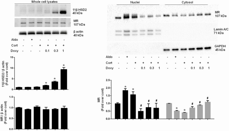
Figure 5.
Nuclear translocation of the mineralocorticoid receptor (MR) in CHO-rMR-pBM14-pTAT3-Gluc.tet-inducible-r11βHSD2 cells induced by 10-nM aldosterone or corticosterone for 1 hour. Cells were lysed and the nuclear and cytosol fractions separated by centrifugation. Laminin A/C and glyceraldehyde-3-phosphate dehydrogenase (GAPDH) are markers for nuclei and cytosol, respectively. *P less than .05 vs no treatment control, and #P less than .05 vs corticosterone, without doxycycline.

