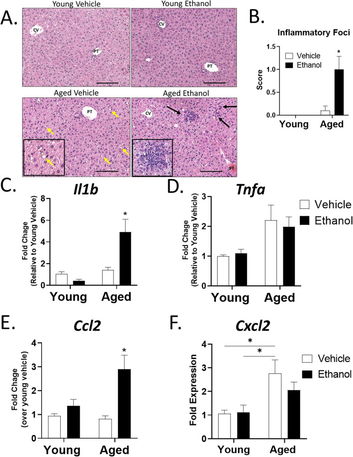
Fig. 3.
Moderate ethanol exposure in aged mice leads to increased hepatic inflammation and pro-inflammatory gene expression. A Representative H&E staining (200x) of livers from young and aged mice given vehicle or ethanol showing dilated sinusoids (yellow arrows and inset representing magnification of the upper right yellow arrow) in aged mice and increased foci of inflammatory cells (black arrows and inset showing magnification of the upper left black arrow) in aged mice given ethanol. Scale bars, 100 µM. PT = portal tract, CV = central vein. B Quantitative scoring of average number of inflammatory foci per 200x field for each group (0 = no foci, 1 = 1–2 foci, 3 = 3–4 foci). n = 4 per group and is representative of 3 individual experiments. C-F. Quantitative RT-PCR of pro-inflammatory and chemokine gene expression from the livers of mice with Hmbs as the endogenous control. Data are shown as mean fold change ± SEM relative to the young vehicle group unless otherwise indicated. n = 6 per group; *p < 0.05 from all other groups, #p < 0.05 from young groups by two-way ANOVA with Tukey’s multiple comparisons test

