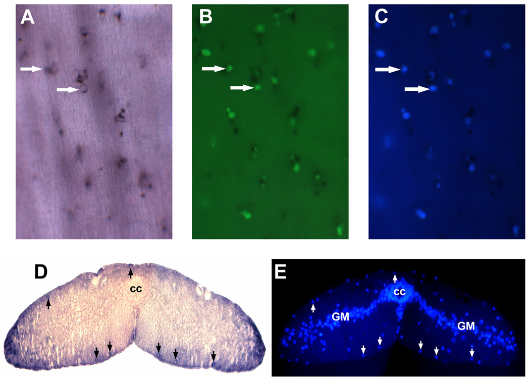
Figure 4. Identification of non-neuronal NT-expressing cells after spinal cord transection.
A–C, The same NT-expressing cells (revealed by wholemount in situ hybridization; white arrows, panel A) were also labeled with IB4 lectin (white arrows in B) and DAPI(white arrows in C); D and E showed that NT-expressing cells were located close to the spinal cord surface (black arrows in D) and were stained simultaneously with DAPI (white arrows in E) that excluded possibility of non-specific NT staining of non-cellular structures. Spinal gray matter (GM). Central canal (cc).

