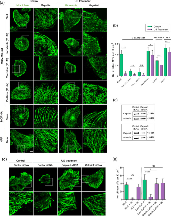
FIGURE 2.

Ultrasound disrupts microtubules by activating calpains in tumor cells. (a) Representative images illustrating microtubule assembly in tumor cells (MDA‐MB‐231) and normal cells (MCF10A and HFF) in the presence of different microtubule targeting agents with and without US treatment, scale bar: 10 μm. (b) Bar diagram demonstrating number of intact microtubules in the defined area in cells under different experimental conditions. n > 30 cells, data are representative of two independent experiments. (c) Western blots showing calpain level in control siRNA‐ and calpain siRNA‐treated tumor cells. (d) Panels displaying microtubule assembly in control siRNA and calpain siRNA treated tumor cells with and without US treatment. (e) Bar diagram demonstrating number of intact microtubules in the defined area under different experimental conditions. Data are representative of two independent experiments, n > 25 cells. *p < 0.05, **p < 0.01, ****p < 0.0001. In all image panels, scale bar: 10 μm
