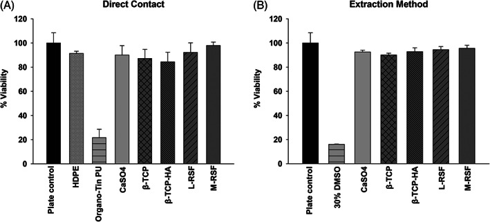
FIGURE 1.

Cytotoxicity assay: (a) Direct contact method: L929 cells (10,000 cells/well) were seeded on 96‐well plate and incubated for 24 h at 37°C, 5% CO2 atmosphere. After 24 h of incubation, media were replaced and scaffolds were placed in direct contact with cells (calcium sulphate [CaSO4], beta tricalcium phosphate [β‐TCP], beta tricalcium phosphate with hydroxyapatite [β‐TCP‐HA], lyophilized‐regenerated silk fibroin [L‐RSF] scaffold, and microparticle‐regenerated silk fibroin [M‐RSF]), high‐density polyethylene (HDPE) (negative control) and Organo‐tin PU (positive control). Plates were incubated further for 24 h. (b) Extraction method: L929 cells were seeded as described above. After 24 h of incubation cells, media were replaced with complete medium (negative control), 30% DMSO (positive control) and extract of all test and control scaffolds. Cells were further incubated for 24 h. After 24 h of treatment MTT proliferation assay was performed and % viability was calculated. Data from three independent experiments are represented
