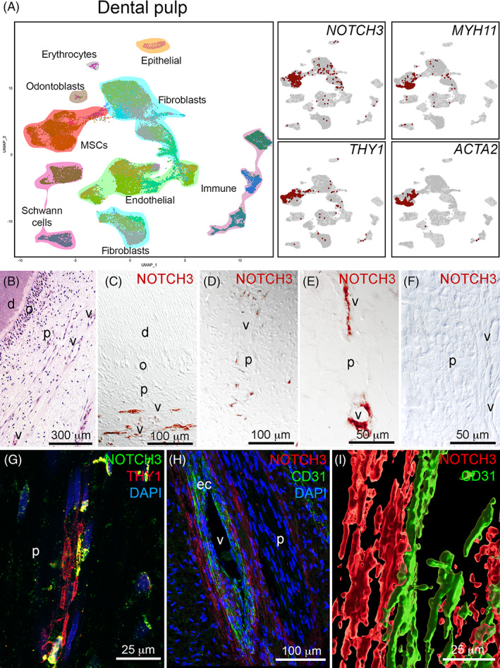
FIGURE 1.

Expression of NOTCH3 in the human dental pulp. A, UMAP visualization of color‐coded clustering of the dental pulp (n > 32 000 cells).37 B, Hematoxylin‐eosin staining showing the structure of the dental pulp. C‐E, Immunohistochemistry against NOTCH3 (red color) in the human dental pulp showing the localization of NOTCH3 in pericytes. F, Negative control for the NOTCH3 staining. G, Immunofluorescent staining showing localization of NOTCH3+ (green color) and THY1+ (red color) mesenchymal stem cells (MSCs). Blue color: DAPI. H, Immunofluorescent staining in the human dental pulp showing NOTCH3 staining in the pericytes (red color) and CD31 staining in endothelial cells (green color). Blue color: DAPI. I, Surface rendering of the relative localization of NOTCH3+ MSCs (red color) and CD31+ endothelial cells (green color). d, dentin; ec, endothelial cells; o, odontoblasts; p, pulp; v, vessels. Scale bars = B = 300 μm; C and D = 100 μm; E and F = 50 μm; G = 25 μm; H = 100 μm; I = 25 μm
