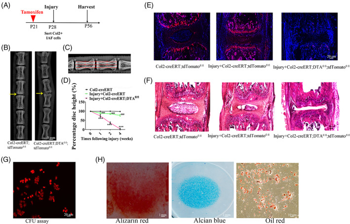
FIGURE 7.

Col2+ progenitors are important for intervertebral disc (IVD) repair. A, The illustration of the experiment design. B, Representative of X‐ray images in Col2‐creERT;tdTomato and Col2‐creERT;DTAfl/fl mice. Yellow arrow: the injured IVD in each group. Scale bar = 5 mm. C, Measurement protocol for the determination of the DHI calculated as [(d + e + f)/(a + b + c + g + h + i) ×100%]. D, DHI at different time‐points following injury. Values are expressed as the mean ± SD (n = 10). DHI, disc height index (n = 6 mice per condition from three independent experiments). Data are mean ± SD. E, Representative fluorescent images of coronal sections of IVD in intact Col2‐creERT;tdTomato, injured Col2‐creERT;tdTomato, and Col2‐creERT;DTAfl/fl;tdTomato mice. Scale bar = 20 μm. F, Representative H&E images of coronal sections of IVD in intact Col2‐creERT, injured Col2‐creERT and Col2‐creERT;DTAfl/fl mice. Scale bar = 20 μm. G, CFU‐F assay in Col2+ IAF progenitor. Scale bar = 20 μm. H, Trilineage differentiation of Col2+ progenitors. Osteogenic (left), chondrogenic (center), and adipogenic (right) differentiation conditions. Statistical significance was determined by one‐way ANOVA and Student's t test. *P < .05, **P < .01, ***P < .0001. NS, not statistically significant. Scale bar = 10 μm
