
FIGURE 5.
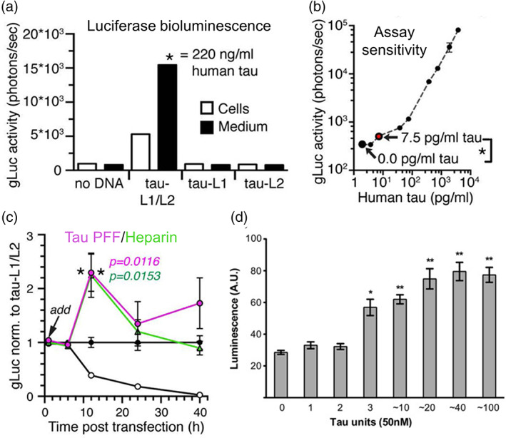
Characterization of split luciferase complementation (SLC) biosensors and their ability to measure the minimum tau units to induce aggregation. (a) Split Gaussia luciferase (split‐gLuc) complementation biosensor illustrating increased complementation of luciferase bioluminescence both in human embryonic kidney 293 (HEK293) cells expressing the split gLuc plasmids (tau‐L1/L2) and in the culture medium where tau oligomers are released by the transfected cells.89 (d) Linear correlation between split‐gLuc activity and tau concentration indicating an assay sensitivity of 7.5 pg/ml tau‐L1/L2 equivalent to 0.16 nM full‐length tau as characterized by human total tau ELISA.89 (c) Treatment of tau preformed fibrils (PFF) and heparin to split‐gLuc biosensor accelerates oligomer formation in cell culture medium after 12 h with a subsequent decrease in the luciferase activity after 24 h.89 (d) Treatment of tauRD oligomers with a minimum of 3 units to HEK293 cells expressing RD‐Nluc/Cluc increases click beetle green luciferase signals.94 Permissions obtained from References 89 and 94