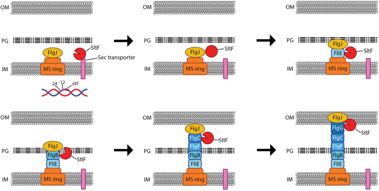
FIG 7.
Model for the possible role of SltF and FlgJ during rod assembly. The scheme shows the internal membrane (IM), the peptidoglycan layer (PG), and the outer membrane (OM). Arrows indicate the sequence of the events taking place during the assembly of the rod. The rod proteins and FlgJ are secreted to the periplasmic space via the flagellar export apparatus and might remain associated with the growing structure, while SltF is secreted through the Sec pathway. Once in the periplasmic space, SltF can recognize the rod proteins and FlgJ. Interaction with FlgJ inhibits its hydrolytic activity (closed red circle, second panel), and interaction with FlgB promotes its activity (open circle, fourth panel). These interactions ensure that the lytic enzyme is under spatial and temporal control and modulate its enzymatic activity in response to the progress of the growing rod.

