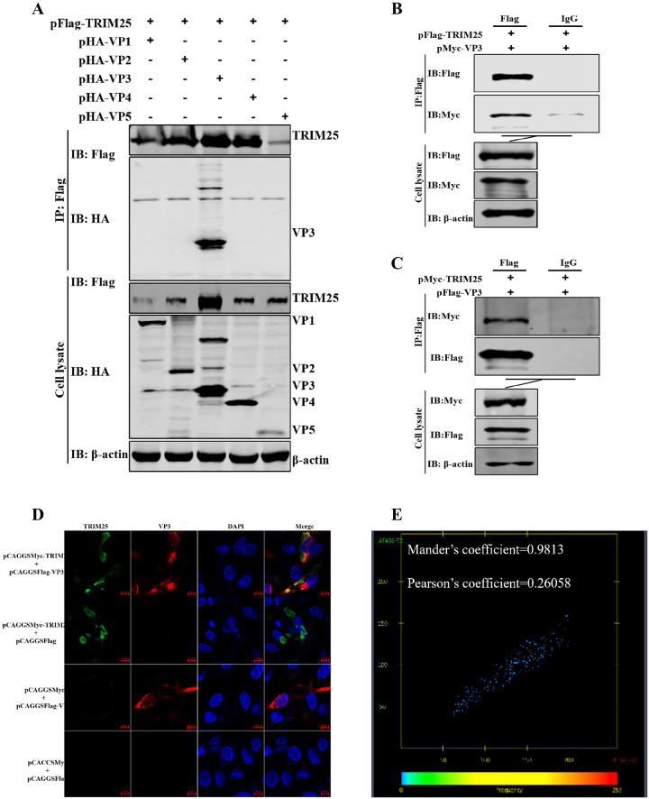
Fig 4. TRIM25 interacts with VP3.
(A-C) Relationship between TRIM25 and VP3 was determined by Co-IP assays in DF-1 cells. (A) Lysates were incubated with anti-Flag mAb and tested using the indicated antibodies by Western blotting. Cells were co-transfected with pFlag-TRIM25 and pHA-VP1, pHA-VP2, pHA-VP3, pHA-VP4, or pHA-VP5 for 48 h, respectively. (B) Cells were harvested after co-transfection with pFlag-TRIM25 and pMyc-VP3 for 48 h. The lysates were incubated with 1 μg anti-Flag mAb or IgG produced in mice. (C) Cells were co-transfected with pFlag-VP3 and pMyc-TRIM25 at 48 h p.i. The lysates were incubated with 1 μg anti-Flag mAb or IgG produced in mice. (D) Confocal assays were used to assess the colocalization between TRIM25 and VP3. DF-1 cells were co-transfected with pFlag-TRIM25 and pHA-VP3 for 36 h. Cells were incubated with anti-Flag mAb produced in mice and anti-HA mAb produced in rabbits and the interaction between TRIM25 and VP3 was determined by confocal assay. (E) Colocalization analysis with Mander’s coefficient (0.9813) and Pearson’s coefficient (0.26058) on the confocal image.

