
FIGURE 2.
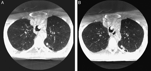
Example images subsequently obtained from low-dose (A) and normal-dose (B) imaging protocols for navigating towards a 6×8 mm lesion found in the apical segment of the left lower lobe. As can be seen, artifacts alike the classically known streaking can be seen more strongly in the low-dose protocol. Although difficult to appreciate on still images, there is also a further difference in image quality for, that is, allowing recognition of the smallest of bronchi that can be navigated to. Note the minor bleeding lateroposterior to the lesion on the image (B) after having performed an initial biopsy.