. 2021 Aug 13;10:e66393. doi: 10.7554/eLife.66393

Figure 4. Plin3 labels nascent lipid droplets and is replaced by a coat of Plin2, which remains with the droplets for the remainder of their lifetime.

(A) Representative confocal micrographs of Fus(plin3-RFP)/+, Fus(EGFP-plin2)/+, or Tg(fabp2:EGFP-PLIN2)/+ fish fed a high-fat meal containing either green or red BODIPY C12 fatty acid analog. Fish were fed for a total of 90 min and chased at room temperature for 30 hr after the onset of the meal. Scale = 20 µm. See also Figure 4—video 1, Figure 4—video 2, Figure 4—video 3. (B) Perilipin fluorescence associated with the lipid droplets and in the cytoplasm was assessed by first segmenting the lipid droplets based on the BODIPY C12 signal, the segmented regions were expanded by three pixels in all dimensions (360 nm) to create a mask which was then applied to corresponding Plin fluorescent image. Mean fluorescence intensity was assessed both in the masked regions (LDs) and outside (cytoplasm). (C–E) Mean fluorescence intensity of Plin3-RFP (C), EGFP-Plin2 (D), and over-expressed human EGFP-PLIN2 (E) in the cytoplasm and associated with lipid droplets over time following a high-fat meal (mean ± SD). Data represent two experiments per genotype, each experiment contained larvae from two clutches (n = 9–12 (C), 4–18 (D), and 3–15 (E) fish per time-point). Due to the complexity and length of the time-course, the 0–8.5 and 16–30 hr time-points represent data from different clutches. The linear correlation coefficients (R) for the 0.75–8.5 hr time-points are –0.3266 (y = –15.20x + 364), 0.7732 (y = 48.91x + (–34.40)), and 0.7277 (y = 52.33x + 36.64) for Plin3, Plin2, and PLIN2, respectively. For the 16–30 hr time-points, the R coefficients are 0.0349 (y = 0.3730x + 180.6), –0.6321 (y = –31.84x + 1113), and –0.2865 (y = –13.33x + 693.8) for Plin3, Plin2, and PLIN2, respectively. (F) Data from C, D, and E were normalized based on the overall mean fluorescence from all individual data points for each genotype and plots were overlaid to better show the relationship between genotypes. A LOWESS local regression line was applied to the normalized data-sets for visualization purposes only.

Figure 4—source data 1. Source data associated with Figure 4C–F.

Figure 4.

Figure 4—figure supplement 1. Tg(fabp2:EGFP-PLIN2) fish over-express human PLIN2 in the yolk syncytial layer and intestine.

Figure 4—figure supplement 1.

(A) Representative images of wild-type and transgenic Tg(fabp2:EGFP-PLIN2)/+ fish at 3 days post fertilization (dpf) highlight EGFP-PLIN2 expression in the yolk syncytial layer. Scale = 500 µm. (B) At 6 dpf, EGFP-PLIN2 is only present in transgenic fish fed a high-fat meal. Fish were imaged ~6.5 hr after the onset of feeding. Scale = 500 µm. (C) Confocal micrographs showing EGFP-PLIN2 decorates lipid droplets containing BODIPY C12 558/568 in the intestine following a high-fat meal. Scale = 10 µm. (D and E) Measurements of standard length (D) and mass (E) at 6 months of age reveal no differences between Tg(fabp2:EGFP-PLIN2)/+ fish and wild-type (WT) siblings. Unpaired t tests, n.s. = not significant, p = 0.6318 (length), p = 0.5604 (mass); n = 42 WT fish and 49 Tg(fabp2:EGFP-PLIN2)/+ fish. No significant differences between genotypes were noted when males and females were compared separately.
Figure 4—figure supplement 1—source data 1. Source data associated with Figure 4—figure supplement 1D and E.
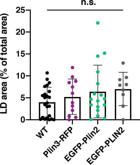
Figure 4—figure supplement 2. Remaining LD area at 30 hr.

Figure 4—figure supplement 2.

The remaining area occupied by BODIPY-positive lipid droplets at 30 hr after the onset of the meal. Mean ± SD, n = 23 WT, 12 Plin3, 17 Plin2, 9 PLIN2, Kruskal-Wallis test, p = 0.2922.
Figure 4—figure supplement 2—source data 1. Source data associated with Figure 4—figure supplement 2.
Figure 4—video 1. Plin3-RFP and BODIPY C12 time series.
Download video file (2.2MB, mp4)
Representative confocal micrographs of Fus(plin3-RFP)/+ fish fed a high-fat meal containing green BODIPY FL-C12 fatty acid analog. Fish were fed for a total of 90 min and chased at room temperature for 30 hr after the onset of the meal. Images correspond to those in Figure 4A. Scale = 20 µm.
Figure 4—video 2. EGFP-Plin2 and BODIPY C12 time series.
Download video file (1.5MB, mp4)
Representative confocal micrographs of Fus(EGFP-plin2)/+ fish fed a high-fat meal containing red BODIPY 558/568-C12 fatty acid analog. Fish were fed for a total of 90 min and chased at room temperature for 30 hr after the onset of the meal. Images correspond to those in Figure 4A. Scale = 20 µm.
Figure 4—video 3. EGFP-PLIN2 and BODIPY C12 time series.
Download video file (1.5MB, mp4)
Representative confocal micrographs of Tg(fabp2:EGFP-PLIN2)/+ fish fed a high-fat meal containing red BODIPY 558/568-C12 fatty acid analog. Fish were fed for a total of 90 min and chased at room temperature for 30 hr after the onset of the meal. Images correspond to those in Figure 4A. Scale = 20 µm.