Figure 2.

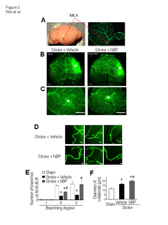
NBP increased collateral growth in the ischemic cortex. (A) The whole brain photo and the GFP green fluorescence image show the middle cerebral artery (MCA) and the surface vasculature distributions in the mouse brain, used to measure MCAO branches at 3 days after stroke. (B) and (C) Representative images showing main and collateral arteries in the ipsilateral hemisphere. Images show the surface of the cortical area and the ischemic core region (*). Bar=1 mm. (D) Enlarged images of collateral arteries in the peri-infarct region. The diameter of collaterals was measured 14 days after stroke. Bar= 20 μm. (E) and (F) Quantified summary of the image analysis of the number and diameter of collaterals. The vessels over the surface were examined and compared. The ischemic insult caused an increase in the number and diameter of the collateral artery. Stroke animals received NBP treatment exhibited even greater increases in the arterial number and diameter. One-way ANOVA; F(2,21)=0.4134 for branching degree I, F(2,21)=38.62 for II, F(2,21)=3.988 for IV, and F(2,10)=21.27 for the diameter, *p<0.05 vs. sham, # p<0.05 vs. stroke + vehicle. N=3 in sham group, N=5 in stroke + vehicle group and stroke stroke + NBP group.
