Figure 1.
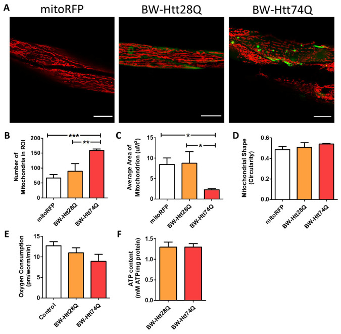
Mitochondrial networks are disrupted in C. elegans models of Huntington’s disease. Worms expressing an expanded, disease-length polyglutamine tract of 74Q in body wall muscle (BW-Htt74Q worms) exhibit mitochondrial fragmentation and mitochondrial network disorganization (see Supplementary Fig. 2). In contrast, worms expressing a shorter, unaffected-length polyglutamine tract of 28Q (BW-Htt28Q worms) have tubular mitochondria, similar to control worms (mitoRFP worms) (A). Mitochondria are labelled with RFP (red), while Htt is labelled with GFP (green). mitoRFP strain is syIs243[Pmyo-3::TOM20:RFP]. BW-Htt28Q and BW-Htt74Q worms also express syIs243[Pmyo-3::TOM20:RFP] transgene. The images shown are from a single focal plane collected on a confocal microscope. Scale bars indicate 15 µM. Quantification of mitochondrial morphology at day 1 of adulthood reveals that BW-Htt74Q worms have an increased number of mitochondria (B) and decreased average mitochondrial area (C), both of which are consistent with increased mitochondrial fragmentation. Mitochondrial shape is not significantly changed in BW-Htt74Q worms compared to BW-Htt28Q and mitoRFP control worms (D). Despite the disruption of mitochondrial morphology, whole worm oxygen consumption (E) and ATP levels (F) are unchanged in BW-Htt74Q worms. A minimum of three biological replicates were performed. Bars indicate the mean value. One-way ANOVA was used to assess significance. Error bars indicate SEM. ROI - region of interest. *p<0.05, **p<0.01, ***p<0.001.

