Figure 2.

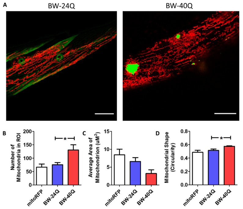
Mitochondrial networks are disrupted in BW-40Q worm model of Huntington’s disease. Worms expressing an expanded, disease-length polyglutamine tract of 40Q in body wall muscle (BW-40Q worms) exhibit mitochondrial fragmentation and disorganized mitochondrial networks (see Supplementary Fig. 4). In contrast, worms expressing a shorter, unaffected-length polyglutamine tract of 24Q (BW-24Q worms) have tubular mitochondria, similar to control worms (mitoRFP worms) (A). Mitochondria are labelled with RFP (red), while polyglutamine protein is labelled with YFP (green/yellow). BW-24Q and BW-40Q worms express syIs243[Pmyo-3::TOM20:RFP] transgene. The images shown are from a single focal plane collected on a confocal microscope. Scale bars indicate 15 µM. Quantification of mitochondrial morphology at day 1 of adulthood reveals that BW-40Q worms have an increased number of mitochondria (B) and a trend towards decreased average mitochondrial area (C). Mitochondrial circularity is significantly increased in BW-40Q worms compared with wild-type worms (D). A minimum of three biological replicates were performed. Bars indicate the mean value. One-way ANOVA was used to assess significance. Error bars indicate SEM. ROI - region of interest. *p<0.05.
