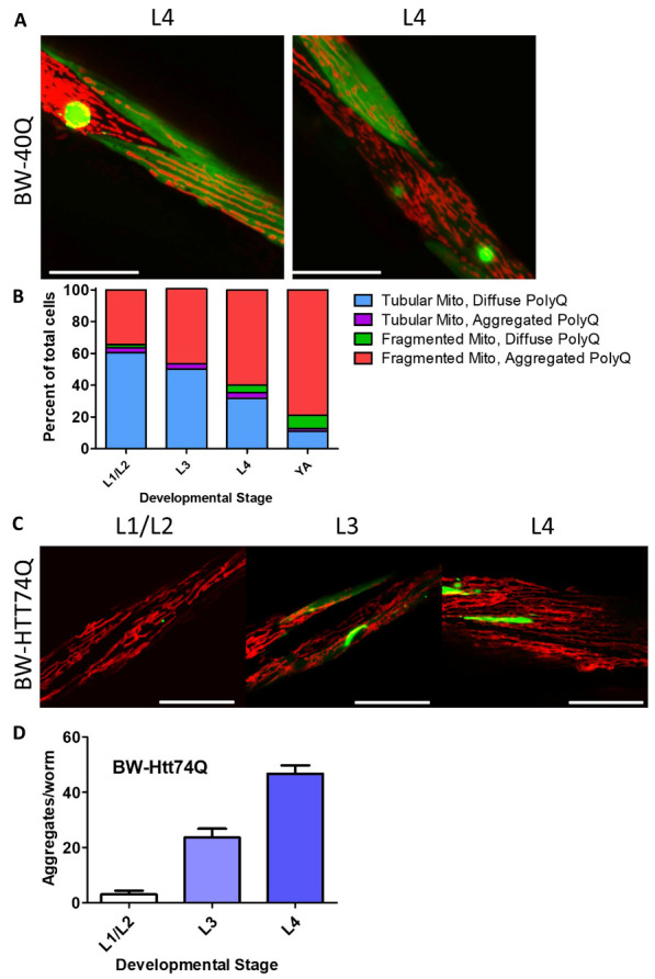
Figure 3.

Disruption of mitochondrial network is associated with polyglutamine aggregation. Images show neighboring body wall muscle cells from worms expressing a disease-length polyglutamine tract in body wall muscles (BW-40Q worms) at the L4 stage of development. One cell has diffuse expression of the polyglutamine (PolyQ) protein and normal, tubular mitochondrial networks, while the other cell has a polyglutamine aggregate and a disrupted mitochondrial network (A). Initially, body wall muscle cells have tubular mitochondria and diffuse polyglutamine protein. Over time, an increasing number of body wall muscle cells have fragmented mitochondria and aggregated polyglutamine. Very few cells exhibit mitochondrial fragmentation and diffuse polyglutamine protein expression, or tubular mitochondria with aggregated polyglutamine protein (B). In BW-Htt-74Q worms, mitochondrial morphology is similar to that in wild-type worms during early development (C). Polyglutamine protein aggregation increases throughout development in BW-Htt74Q worms (D). The images in panels A and C are compressed z-stacks collected on a confocal microscope. Scale bars indicate 25 µM. A minimum of three biological replicates were performed. Bars indicate the mean value. Error bars indicate SEM.
