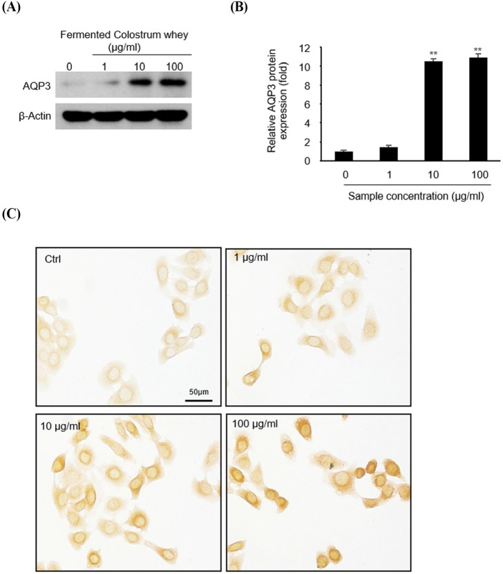
Fig. 3. Effect of fermented colostrum whey on AQP-3 protein expression.
(A) HaCaT cells were treated with different amounts (0, 1, 10, and 100 μg) of fermented colostrum whey for 3 h. AQP-3 protein expression was analyzed by immunoblotting. The β-actin antibody was used as a control to confirm that equal amounts of each protein were loaded. (B) In the bar graph, the relative intensity of AQP-3 is expressed as a ratio of AQP-3/β-actin and compared with that for the untreated group. Data were presented as the means±SE values of three independent experiments (n=3). (C) AQP-3 protein expression was analyzed by immunocytochemistry at a 400× magnification (Scale bars=50 μm; all images were taken at the same magnification). Immunocytochemical staining was performed using the DAB solution.

