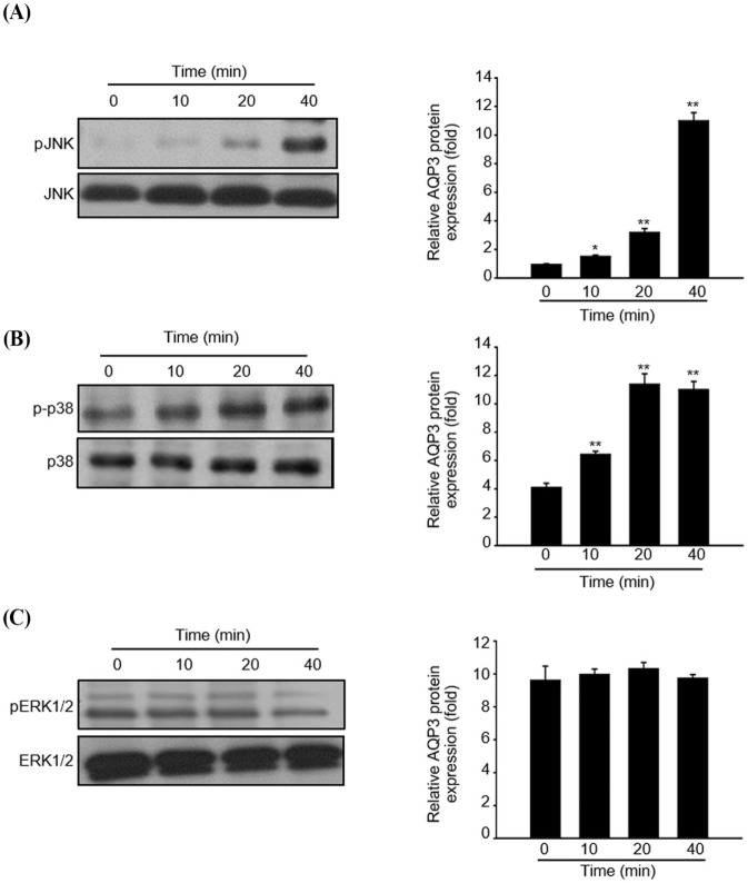
Fig. 4. Role of MAPK signaling in fermented colostrum whey-induced proliferation of HaCaT keratinocytes.
HaCaT cells were treated with 100 μg/mL of fermented colostrum whey for varying time periods. Phosphorylation levels of MAPKs such as (A) p-JNK, (B) p-p38, and (C) pERK1/2 were analyzed by western blotting. The β-actin antibody was used as an internal control to confirm that equal amounts of each protein were loaded. The bar graph represents the normalized values of the densities of each band relative to the densities of the bands of the non-active form of each protein. The values represent the mean±SE values of three independent experiments (n=3). JNK, Jun N-terminal kinase. MAPK, mitogen-activated protein kinase.

