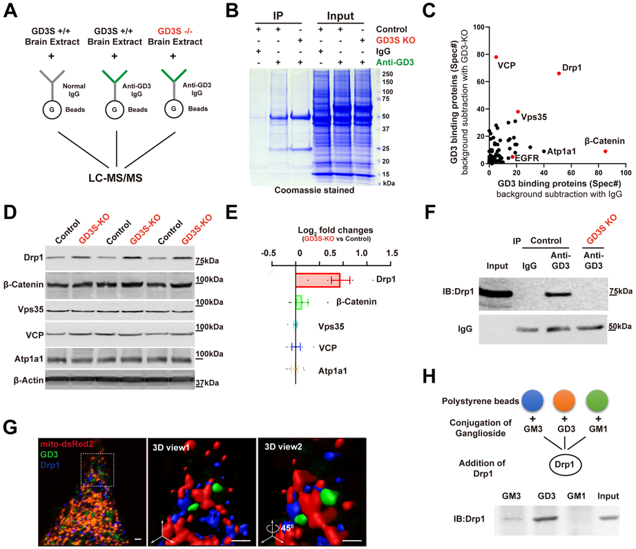
Figure 5. Identification of Drp1 as a GD3-binding protein.

(A) Proteomic workflow for the identification of GD3-interacting proteins. (B) Representative Coomassie-stained SDS gel of proteins bound to bead-coupled anti-GD3 antibody incubated with brain extracts, with indicated negative control. (C) Frequency of detection of all peptides (total spectra count) for proteins identified. (D) Lysates (30 μg protein) of DG were immunoblotted with the indicated antibodies. (E) Quantification of data from (D) (normalized by control). n = 6 mice / group. (F) Representative Western blot analyses for the presence of immunoprecipitates. (G) Representative 3D images showing the co-localization of endogenous GD3 and Drp1 on mitochondria in NSCs with mito-DsRed2 and immunostained with anti-Drp1 and mAb R24. Boxed area in the left image was enlarged and viewed at different angles. Scale bars, 1 μm. (H) Interaction of GSL-coated polystyrene beads with Drp1.
