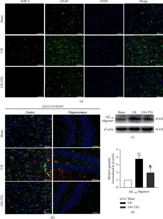
Figure 4.

Intervention effects of TXL on AQP-4 polarization loss and Aβ accumulation after reperfusion. (a) Representative pictures of the double immunofluorescence staining of AQP-4 (red) and GFAP (green) in ischemic cortex; scale bars, 50 μm. (b) Representative pictures of the double immunofluorescence staining of Aβ (red) and GFAP (green) in ischemic cortex and hippocampus; scale bars, 50 μm. (c-d) Protein levels of Aβ1–42 oligomers in each group; n = 6. Data are presented as mean ± SEM. ∗∗P < 0.01 versus the sham group. ▲P < 0.05 versus the I/R group.
