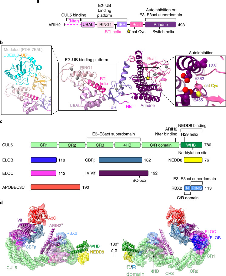
Fig. 1.
a, ARIH2 schematic color-coded by domains. The catalytic Cys310 (cat Cys) in the Rcat domain is indicated with a yellow star. Nter, N terminus. b, Crystal structure of autoinhibited ARIH2 (residues 51–493) is shown in center, with domains colored as in a, zinc atoms as spheres and Ariadne domain helices numbered. The ARIH2 UBAL, RING1, RTI helix and IBR domains form an E2~UB-binding platform. Left inset, UBE2L3~UB (from complex with neddylated CRL1-bound ARIH1, ref. 23) modeled onto the ARIH2 E2~UB-binding platform. Right inset, close-up (rotated 30° in x and 30° in y) highlighting L381, E382 and E455 mediating autoinhibition. ARIH2 catalytic Cys thiol is shown as a yellow sphere. c, Color-coded schematics of subunits and domains of neddylated CRL5Vif-CBFβ and APOBEC3C (A3C). d, Model of ARIH2* (full-length ARIH2 with L381A, E382A and E455A residue substitutions) complex with neddylated CRL5Vif-CBFβ and A3C in cryo-EM reconstruction low-pass filtered to 7.5 Å. Coordinates for ARIH2*, RBX2 and a portion of neddylated CUL5 (CR3 domain through the C terminus) were built into the map shown in Extended Data Fig. 2b. Structures of A3C39 and Vif-CBFβ-ELOBC-CUL5 N-terminal domain36 were fit in density.

