Fig. 2.
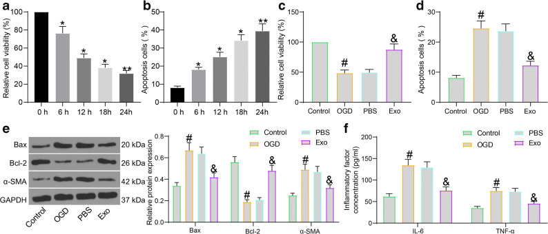
Exo treatment protects cardiomyocytes against OGD-induced damage. a Viability of cardiomyocytes at different time points after OGD treatment determined by the MTT assay; b Apoptosis of cardiomyocytes at different time points after OGD treatment examined by flow cytometry; c Viability of cardiomyocytes after Exo treatment examined by the MTT assay; d Apoptosis of cardiomyocytes after Exo treatment detected by flow cytometry; e Protein levels of apoptosis-related factors (Bax and Bcl-2) and the fibrosis-related factor α-SMA in OGD- and Exo-treated cardiomyocytes quantified by western blot analysis; f Production of pro-inflammatory cytokines (IL-6 and TNF-α) in OGD- and Exo-treated cardiomyocytes determined using ELISA kits. Data were collected from three experiments and exhibited as mean ± SEM. Differences were analyzed by one-way ANOVA (a–d) or two-way ANOVA (e, f); *p < 0.05, **p < 0.01 vs. 0 h; #p < 0.05 vs. control group; &p < 0.05 vs. PBS group.

