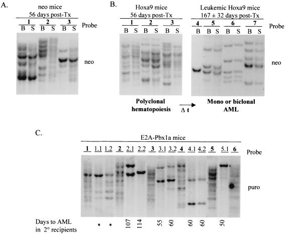
FIG. 7.
Southern blot analysis of DNA isolated from bone marrow of primary neo (A) and Hoxa9 (B) mice and primary and secondary E2A-Pbx1a mice (C). The DNA was digested with EcoRI, which cuts the integrated provirus once, thus generating unique fragments specific for the proviral integration site(s). The membranes were hybridized with a neo-specific probe to detect the neo control and Hoxa9 proviral fragments and a puro-specific probe to detect those of the E2A-Pbx1a provirus. Each primary mouse is identified with specific number in bold, and its secondary recipients are identified with derivatives thereof. All secondary recipients were transplanted with 1 × 10⁶ to 2 × 10⁶ bone marrow or spleen cells from the donor mouse. Where applicable, the time (in days posttransplantation [post-Tx]) for the development of acute leukemia in the secondary E2A-Pbx1a mice is shown. Asterisks denote secondary (2°) mice that developed MPS.

