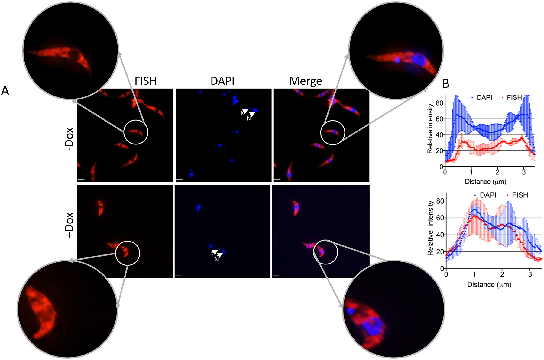
Figure 4. DRBD18 RNAi causes partial accumulation of mRNA in the nucleus.

PF T. brucei cells harboring the DRBD18 RNAi construct were used to assess the relative abundance of mRNA in the nucleus upon depletion of DRBD18. (A) Subcellular localization of total mRNA in uninduced (−Dox) or induced (+Dox) DRBD18 RNAi cells was monitored by fluorescence in situ hybridization (FISH) using fluorescently labelled oligo(dT). DAPI was used to stain the DNA of the nucleus (N) and the kinetoplast (K). An overlay of the FISH and DAPI signals (Merge) is shown. Bars, 3.2 μm. Images of individual cells were enlarged to enhance visualization of the nucleus. (B) Quantification of the fluorescence intensities of FISH (red-Alexa 594) and DAPI (blue) fluorophores were calculated on a line drawn across the nucleus, with overhangs covering the cytosol, using ImageJ (NIH) software. Two biological replicate experiments were performed. The relative intensities represent the average ± SD from six randomly selected cells (three from each replicate).
