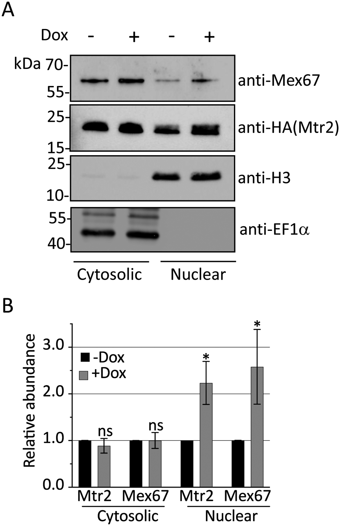
Figure 6. DRBD18 RNAi causes partial accumulation of export receptors in the nucleus.

(A) The relative abundance of Mtr2 and Mex67 in nuclear and cytosolic fractions in uninduced (-Dox) or induced (+Dox) DRBD18 RNAi cells were analyzed by subcellular fractionation followed by Western blot analysis. Anti-Histone H3 (H3) and anti-EF1α antibodies were used as loading controls for nuclear and cytosolic fractions, respectively. (B) Quantification of Western blots in (A). Relative abundance of Mex67 or Mtr2 in nuclear and cytosolic fractions was normalized to the expression of Histone H3 and EF1α, respectively. The normalized protein expression in nuclear and cytosolic fractions from the +Dox were then compared to that of the −Dox (which was set to 1) to calculate the relative abundances in the DRBD18 knockdown. Bar graphs represent the average and standard deviation (SD) of four samples (two biological replicates, each with two technical replicates). Significance was determined by unpaired t-test with Welch’s correction. *p < 0.05 and ns = non-significant.
