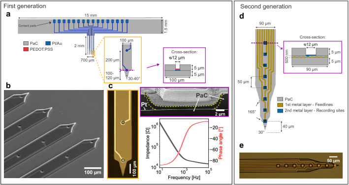
Figure 1.
Overview of the flexible intracortical probes. First generation of devices: (a) Critical dimensions of the Michigan-style arrays consisting of four shanks and four microelectrodes per shank. One shank has a cross-section of 1000 μm. (b) Close-up SEM image of the four shanks with recording sites and metal traces consisting of a metal film sandwiched between two PaC layers. (c) Close-up optical image (left) of the tip of a shank and the PEDOT:PSS coating with its characteristic blue colour. FIB section of electrode coating (right top, PEDOT:PSS bordered in yellow). Bode plot (right bottom) of the spin-coated PEDOT:PSS films on a single electrode array (n = 16 electrodes, mean ± std). Second generation of devices: (d) A double-metal-layer process was utilized to double the number of recordings sites per shank while keeping the shank width and thickness. The layout of the bond pads is comparable to the first generation. (e) Optical image of one shank with eight recording sites.

