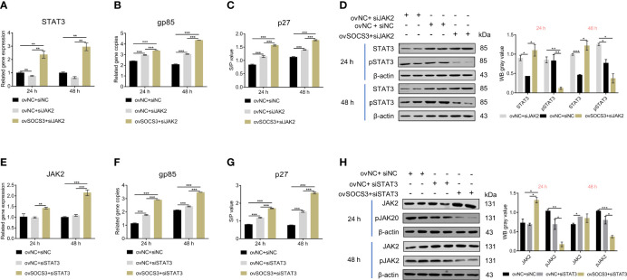
Figure 6.
The effects of JAK2/STAT3 phosphorylation on ALV-J virus replication. The pcDNA3.1-SOCS3 and si-JAK2 were co-transfected into DF-1 cells. The transfected cells were infected with 105 TCID50/mL ALV-J strain SCAU-HN06 after 24 h. The (A) STAT3 gene and (B) ALV-J virus were detected by qRT-PCR. The (C) ALV-J virus (p27) and (D) STAT3 phosphorylation were detected by ELISA and WB, respectively. The pcDNA3.1-SOCS3 and si-STAT3 were co-transfected into DF-1 cells. The transfected cells were infected with 105 TCID50/mL ALV-J strain SCAU-HN06 after 24 h. The (E) STAT3 gene and (F) ALV-J virus were detected by qRT-PCR. The (G) ALV-J virus (p27) and (H) STAT3 phosphorylation were detected by ELISA and WB, respectively. pcDNA3.1+si-NC, ovNC + siNC; pcDNA3.1+si-JAK2, ovNC + si-JAK2; pcDNA3.1-SOCS3 + si-JAK2, ovSOCS3 + siJAK2; pcDNA3.1+si-STAT3, ovNC + si-STAT3; pcDNA3.1-SOCS3 + si-STAT3, ovSOCS3 + si STAT3. These experiments were performed independently at least three times with similar results. Differences in data were evaluated by the Student’s t-test. The error bars are the standard error of the mean (SEMs) (*P ≤ 0.05, **P ≤ 0.01, and ***P ≤ 0.001).

