Figure 5.
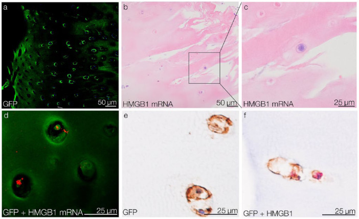
Migration of CPCs into OA tissue and their HMGB1 expression ex vivo. (a) After 5 days of migration, GFP-tagged CPCs are detected in OA cartilage tissue by confocal microscopy. (b) detection of HMGB1 mRNA in migrated CPCs via ISH. (c) Detail of a HMGB1 mRNA-positive migrated CPC. (d) Confocal reflection microscopy confirms HMGB1 mRNA (red) in migrated GFP-tagged CPCs (green). (e) IHC results show that migrated CPCs are GFP-positive. (f) IHC double-staining shows that migrated GFP-positive CPCs (brown) express HMGB1 (red). CPCs = chondrogenic progenitor cells; HMGB1 = high mobility group box 1 protein; OA, osteoarthritis; GFP = green fluorescent protein; ISH = in situ hybridization; IHC = immunohistochemistry

