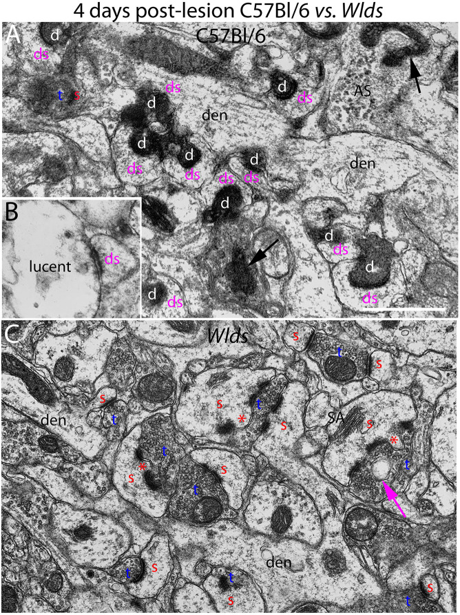
FIGURE 2.
Ultrastructure of synapses at 4 days post-lesion in C57Bl/6 and Wlds mice. (A) Synapses in the middle molecular layer exhibiting typical dark-dense degeneration at 4 days post-lesion in C57Bl/6 mice. Spines contacted by degenerating presynaptic terminals had abnormal morphologies. (B) Presynaptic terminal exhibiting the lucent form of degeneration. (C) Non-degenerating presynaptic terminals in the middle molecular of Wlds mice at 4 days postlesion. Spine heads are large and some have multiple PSDs and some had finger-like protrusions (termed spinules) that extended into the presynaptic terminal (marked by red asterisks). Large complex spine apparatuses (SA) were also evident. Red s, spine head contacted by non-degenerating synapse; Blue t, non-degenerating synaptic terminal; white d, degenerating synaptic terminal; Purple ds, spine contacted by degenerating presynaptic terminal; den, dendrite; ax, myelinated axon. Calibration bar in (A) = 0.25 μm and applies to (A,B).

