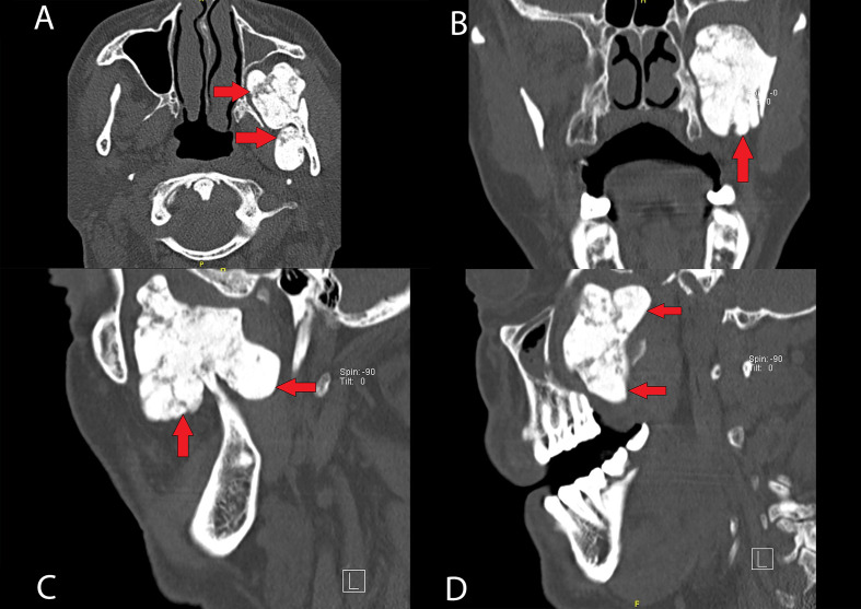
Figure 2.
Cranial CT scan imaging of the mandibular osteoma, different projections: axial (A), coronal (B) and sagittal (C, D). We can see a bony-like density mass, measuring 45×33×50 mm (pointed with arrows) and its anatomical relation with mandibular left condyle, in continuity with the internal cortex, as well as its polylobulated shape (A, C).

