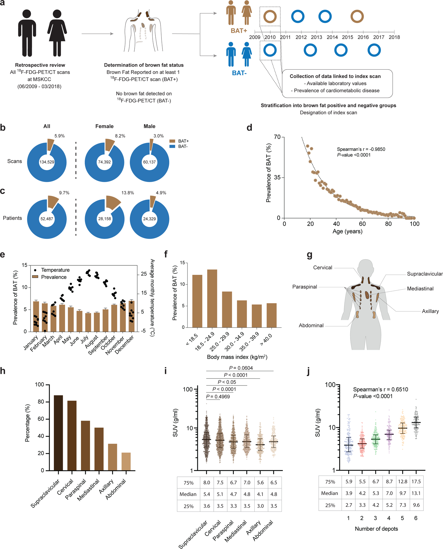
Fig. 1: Study design, association of brown fat with patient demographics and characterization of brown fat activity across adipose depots.

a,18F-FDG PET/CT reports were stratified by presence or absence of brown fat. b, Prevalence of brown fat per 18F-FDG PET/CT scan (n=134,529), further stratified according to 18F-FDG PET/CT scans in female (n=74,392) and male (n=60,137) individuals. c, Prevalence of brown fat in all patients (n=52,487), stratified by female (n=28,158) and male (n=24,329) sex. d, Correlation between brown fat prevalence and age. e, Brown fat prevalence, based on 18F-FDG PET/CT scans, and correlation with outdoor temperature in the month of the scan between 1 June 2009 and 31 March 2018. Bars depict means; error bars are standard deviations. f, Brown fat prevalence and BMI. g, Schematic of brown fat depot location in humans. h, Prevalence of brown fat in defined anatomic locations, derived from all 18F-FDG PET/CT scans with brown fat in 2016 (n=1,091 scans). i, Comparison of BAT activity measured in SUV across the different depots (n=1,091). Compared with supraclavicular depots, BAT activity was significantly lower in paraspinal (P<0.0001), mediastinal (P=0.0185) and axillary (P<0.0001) depots. Activities were compared by Kruskal-Wallis test with Dunn’s post hoc test; all tests were two-sided. Line depicts median; error bars are 25th and 75th percentile. j, Correlation between highest measured brown fat activity per patient and cumulative number of adipose depots with detectable brown fat activity (n=1,091 scans). Line depicts median; error bars are 25th and 75th percentile.
