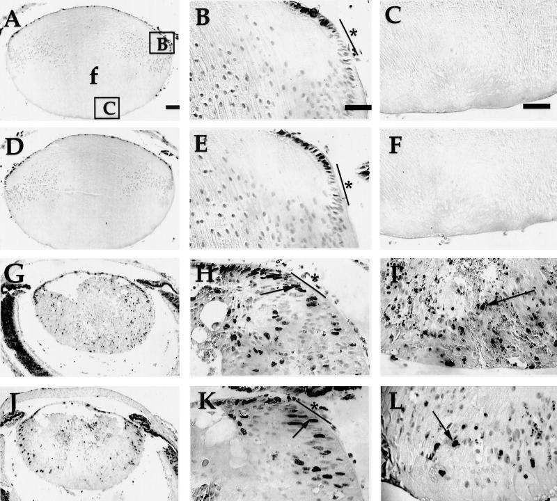
FIG. 2.
In situ detection of proliferation in lenses from neonatal E7 transgenic mice on E2F-1-sufficient or -deficient backgrounds by using a BrdU incorporation assay. BrdU incorporated into newly synthesized DNA for 1 h was detected in paraffin sections (5-μm thickness) of neonatal eyes from nontransgenic (A, B, and C), E2F-1−/− (D, E, and F), E7/E2F-1+/− (G, H, and I), and E7/E2F-1−/− (J, K, and L) mice. Representative sections are shown. Panels B, E, H, and K show higher magnifications of the right equatorial regions of the lenses shown in panels A, D, G, and J, respectively (see box B in panel A). Panels C, F, I, and L show higher magnifications of the posterior regions of the lenses shown in panels A, D, G, and J, respectively (see box C in panel A). No detectable signal was observed in control sections from neonates in which BrdU was not injected (data not shown). Arrows point to dark diaminobenzidene-stained nuclei indicating BrdU-positive nuclei; light nuclei are BrdU-negative nuclei. e, epithelial cells; f, fiber cells; bars and asterisks in panels B, E, H, and K denote approximate boundaries of the transitional zone; the proliferation zone is anterior to the transitional zone. (See Materials and Methods section for a more complete definition of these zones.) Bar, 100 μm for panels A, D, G, and J and 50 μm for other panels. In all panels the anterior of the lens is oriented at the top.

