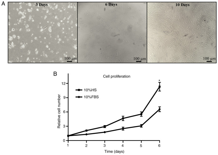
Figure 2.
Isolation and proliferation of Ad-MSCs. Ad-MSCs were observed after isolation from adipose tissue and relative proliferation was assessed under HS and FBS conditions. (A) Cells were evaluated for cell morphology and confluence at 3, 6 and 10 days after isolation. Red blood cells were observed at day 3 as small circular cells. Ad-MSCs displayed adherence to culture flasks, with a fibroblastoid morphology (scale bars, 100 µm). (B) Relative cell proliferation was analyzed for 6 days; the relative proliferation increased significantly with cell culture under HS conditions. *P<0.05 vs. 10% FBS. Ad-MSCs, adipose-derived mesenchymal stem cells; HS, human serum.

