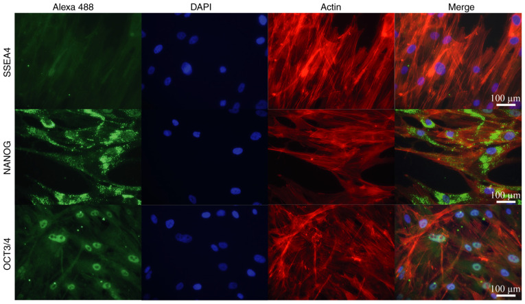
Figure 3.
Immunofluorescence analysis of pluripotent factors in adipose-derived MSCs. MSC pluripotential markers NANOG and OCT3/4 were visualized by secondary antibody conjugated with Alexa 488 (green fluorescence). The negative control SSEA4 did not exhibit a signal in the cells. Nuclei and actin filaments were visualized with DAPI and rhodamine phalloidin, respectively (scale bars, 100 µm). MSCs, mesenchymal stem cells; NANOG, Nanog homeobox; OCT3/4, octamer-binding transcription factor; SSEA4, stage-specific embryonic antigen 4.

