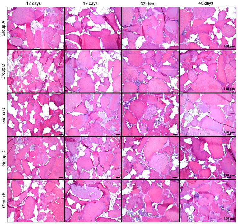
Figure 7.
H&E staining of cellularized biphasic scaffolds. Matrix formation in cellularized biphasic scaffolds subjected to the aforementioned differentiation schemes was analyzed by H&E staining at different times of exposure (12, 19, 33 and 40 days). The generation of a filling tissue may be observed as an amorphous and acellular tissue between cartilage matrix fragments (scale bar, 100 µm).

