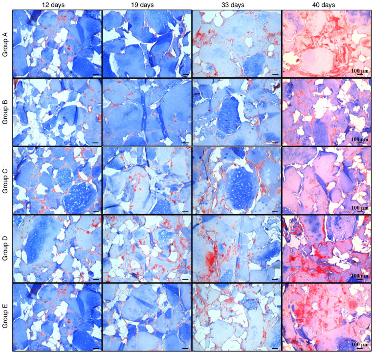
Figure 8.
Masson's trichrome staining of cellularized biphasic scaffolds. Matrix formation in cellularized biphasic scaffolds exposed to the aforementioned differentiation schemes was analyzed by Masson's trichrome staining at different times of exposure (12, 19, 33 and 40 days). Collagen matrix with blue staining and fibrous tissue with red staining may be observed (scale bar, 100 µm).

