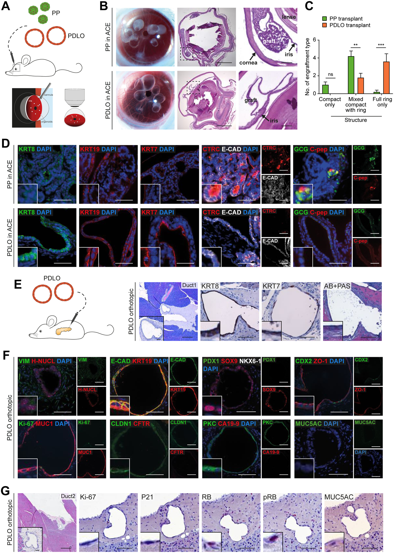
Figure 4. Development of human duct-like tissue after xenotransplantation of PDLOs.

(A) Scheme of transplantation into the anterior chamber of the mouse eye (ACE). (B) Growth of grafted organoids on the iris 5 weeks after transplantation. Left panel: Image of eyes transplanted with PPs or PDLOs. Right panels: HE staining of sagittal section of explanted eyes with PDLO graft on the iris. (C) Quantification of observed engraftment types (Mean±SEM; n=5 mice per group; ordinary two-way Anova with Sidak’s multiple comparison test)(D) IF staining of PP-derived grafts revealed acinar, ductal, and endocrine cells in the ACE, while marker expression of PDLO-derived grafts was restricted to the ductal pancreatic lineage (CTRC, Chymotrypsin C; C-pep, C-peptide). (E) Orthotopic transplantation scheme and HE/IHC images demonstrating engraftment site 8 weeks after transplantation (n=5 mice). (F) PDLO transplants expressed ductal epithelium-specific proteins, MUC1, E-CAD, KRT19, and CLDN1, lost transcription factors, PDX1 and CDX2, but lacked CFTR and CA19-9 expression. (G) WT PDLO transplant stained for proliferation- and cell cycle-related proteins and the dysplastic marker MUC5AC (RB, retinoblastoma protein; pRB, phosphorylated RB). B,E-G: Scale bars: 100 μm. Insets in the corners are 4x enlarged. Except in overview staining: 500 μm, here insets: 50 μm. D: Scale bar: 50 μm, insets are 2x enlarged.
