Fig. 2.
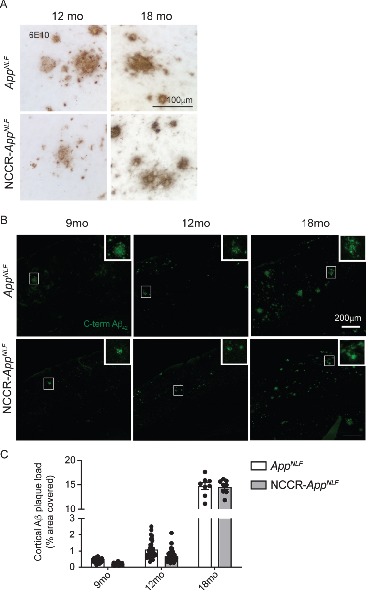
Aβ plaque load in AppNLF KI mice is not altered by neuronal cell cycle re-entry. A) DAB stained Immunohistochemical (6E10 antibody) image showing Aβ deposition in the primary somatosensory areas of cortex in AppNLF KI and NCCR-AppNLF KI mice. Scale bar 100μm. B) Representative immunofluorescence images of C-terminal Aβ42 antibody-positive Aβ plaque staining in the primary somatosensory areas of cortex in AppNLF and NCCR-AppNLF mice at 9, 12, and 18 months of age used for Aβ plaque load analysis. Insert is a magnified image of the region inside the white square. Cortical areas covered by C-terminal Aβ42 positive extracellular signals were quantified in four matched coronal brain sections from each perfusion fixed animals using the Area Fraction Fractionator probe (Stereoinvestigator, MBF). Scale bar 200μm. C) Two-way ANOVA showed significant age effect (F (2, 140) = 3437, p < 0.0001) but no difference between genotypes (F (1, 140) = 3.637, p = 0.0586), 9 months: n = 8/group; 12 months: n = 8-9/group; 18 months: n = 2/group. Bars represent mean±SEM.

