Fig. 4.
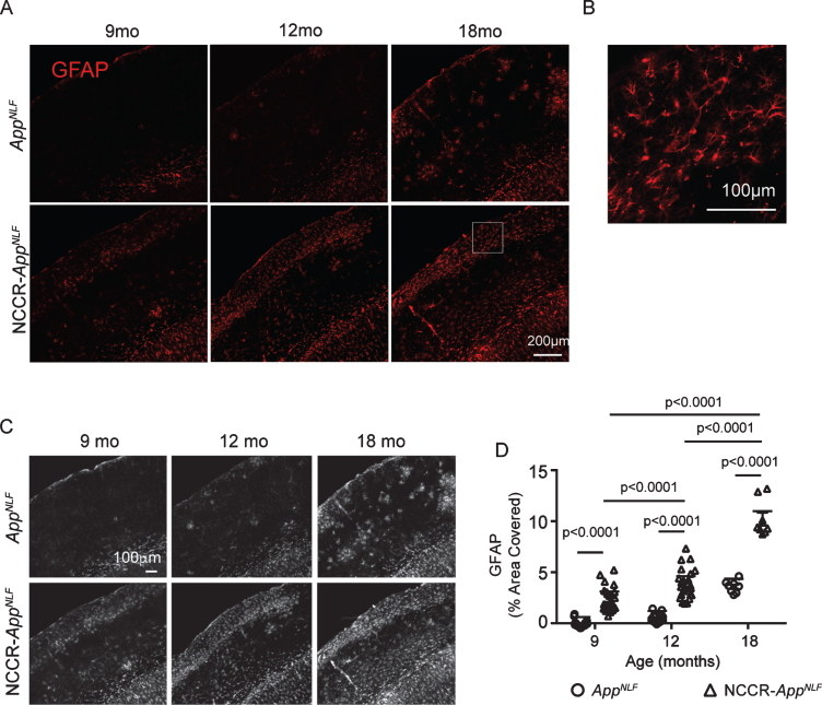
Neuronal cell cycle re-entry increases astrocytosis in AppNLF mice. A) Representative fluorescently labeled images of cortical areas (primary somatosensory region) stained using GFAP antibody. B) Magnification of white inset in (A) demonstrating astrocyte specific GFAP-labeling. C) Grayscale images of GFAP immunofluorescence labeled whole cortical regions in brain sections from 9‐, 12‐, and 18‐month-old NCCR‐AppNLF and AppNLF animals were evaluated using Image J. D) Two-way ANOVA shows significant age effect (F(2, 105) = 196, p < 0.0001), genotype effect (F(1, 105) = 359.3, p < 0.0001), and interaction effect (F(2, 105) = 24.68, p < 0.0001). GFAP immunoreactive cortical area is greater in the NCCR- AppNLF animals compared to age-matched AppNLF animals at all ages. Furthermore, NCCR-AppNLF animals show progressive increase with age (Tukey’s multiple comparison test, 9 months versus 12 months, p < 0.0001; 9 months versus 18 months, p < 0.0001; 12 months versus 18 months, p < 0.0001). Following numbers of animals were examined; at 9 months of age: NCCR-AppNLF, n = 4; AppNLF, n = 4‐ at 12 months of age: NCCR-AppNLF, n = 4; AppNLF, n = 4; at 18 months of age: NCCR-AppNLF, n = 2; AppNLF, n = 2. Four matched coronal sections were measured per animal. Each data point represents a measure from each section.

