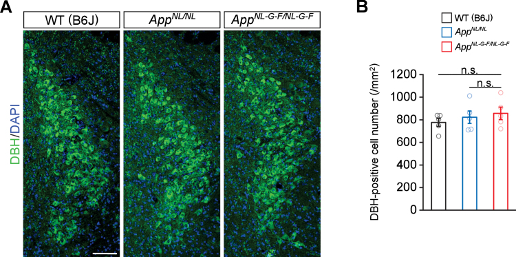
Fig. 2.
No prominent neuron loss is detected in the LC in 24-month-old AppNL-G-F/NL-G-F mice. A) Representative images of the LC from frozen coronal brain sections immunostained with anti-DBH (indicated by green) were shown (blue indicated DAPI staining). B) Number of DBH-positive cells was measured and expressed as cell number per area (/mm2). Scale bar represents 100μm. n = 5/genotype. n.s., not significant.

