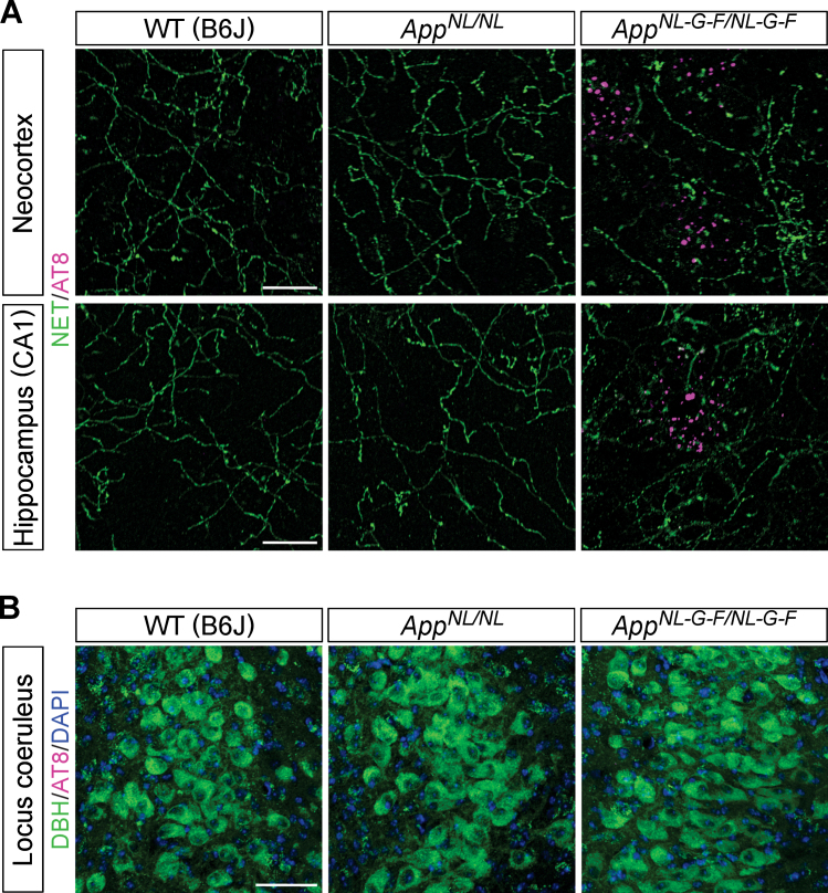
Fig. 3.
Phospho-tau pathology is not associated with the LC-NA system. A) Representative images of the neocortex and hippocampal CA1 subfield from frozen coronal brain sections immunostained with anti-phospho tau (AT8) (indicated by magenta) and anti-NET (indicated by green) antibodies were shown. Scale bars represent 20μm. B) Representative images of the LC from frozen coronal brain sections immunostained with anti-phospho tau (AT8) (indicated by magenta) and anti-DBH (indicated by green) antibodies were shown (blue indicated DAPI staining). Scale bar represents 50μm. n = 3/genotype.

