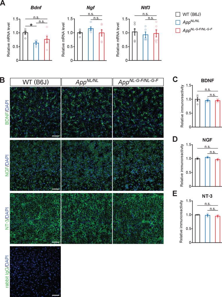
Fig. 7.
No prominent reduction in neurotrophic factors for noradrenergic neurons are detected in the neocortex. A) mRNA levels of Bdnf, Ngf, and Ntf3 in the cortex were analyzed by qRT-PCR. n = 5–6/genotype. *p < 0.05 versus WT (B6J). B) Representative images of the neocortex from frozen coronal brain sections immunostained with anti-BDNF, anti-NGF, and anti-NT-3 antibodies (all indicated by green) were shown. Sections stained with secondary anti-rabbit IgG antibody (indicated by green) was served as a negative control (blue indicated DAPI staining). C–E) Immunoreactivity of BDNF (C), NGF (D), and NT-3 (E) were evaluated and expressed as a relative ratio to WT (B6J). Scale bars represents 50μm. n = 3–5/genotype. n.s., not significant.

