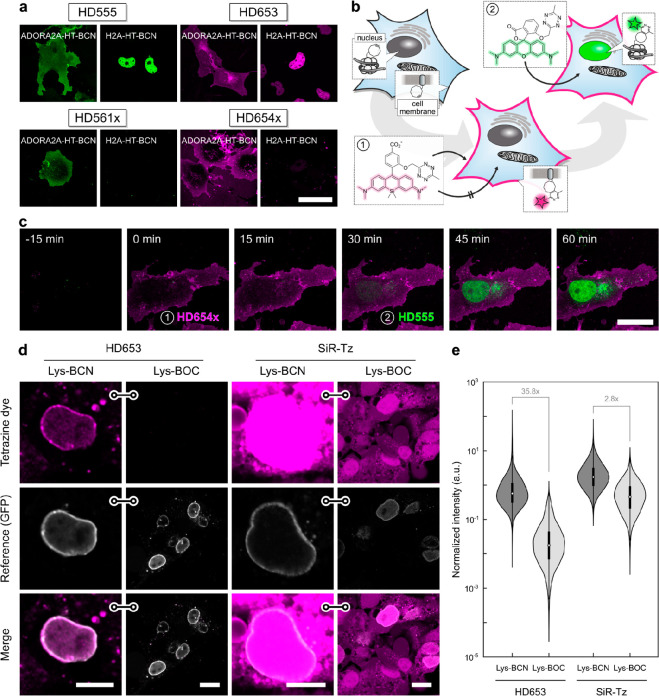
Figure 4.
Wash-free multicolor and unnatural amino acid imaging in live cells. (a) Wash-free labeling of intra- and extracellular targets in COS-7 cells. Cells were transiently transfected with ADORA2A–HaloTag (plasma membrane) or H2A–HaloTag (nucleus), loaded with HTL–BCN, and incubated with HDyes at 1 μM for 30 min. Scale bar 50 μm. For reference and controls, see Figures S16–19. (b) Dual-color labeling with a single chemistry. Intra- and extracellular proteins H2A and ADORA2A are targeted via HaloTag and HTL–BCN and then incubated with cell-impermeable HD654x for 30 min followed by addition of HD555. (c) Time-lapse confocal microscopy shows labeling of intra- and extracellular targets with spectrally distinct fluorophores can be achieved without any washing. Scale bar 20 μm. For individual color channels and reference staining, see (Figure S23). (d) Un-natural amino acid labeling of pEGFPN149TAG–Nup153 with Lys–BCN or Lys–BOC (control) in COS-7 cells. Live-cell confocal microscopy after incubation with tetrazine dyes HD653 and SiR-Tz at 500 nM for 30 min and mere buffer replacement. Scale bars: 10 μm (Lys–BCN) or 20 μm (Lys–BOC). (e) Quantitative comparison of target and background labeling with flow cytometry analysis. pEGFPN149TAG–Nup153 COS-7 cells were loaded with either Lys–BCN for specific or Lys–BOC for unspecific labeling. Intensity of HD653 and SiR-Tz was normalized to EGFP and signal-to-background ratios calculated from population medians.

