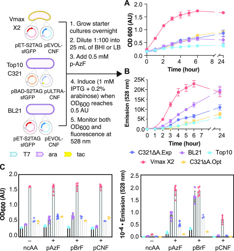
Figure 2.
Growth and sfGFP expression in Vmax X2 versus traditional (Top10, BL21) and genomically recoded (C321)39,40E. coli strains. (A) Plot of the OD600 of each cell growth as a function of time. Vmax X2 and BL21 cells were transformed with pET-S2TAGsfGFP and pEVOL-CNF, whereas Top10 and C321 cells were transformed with pBAD-S2TAGsfGFP and pULTRA-CNF (Top10, C321) to induce expression of sfGFP bearing an ncAA at the second position of sfGFP. After induction, cells were grown for 24 h at 37 °C (Vmax X2, BL21, Top10, C321.ΔA.exp) or 34 °C (C321.ΔA.opt) in the presence of 0.5 mM pAzF. (B) Plot of the emission of each cell growth at 528 nm as a function of time. (C) Plots comparing the OD600 and 528 nm fluorescence of each growth at the 4 h time point in the presence or absence of pAzF, p-bromo-l-phenylalanine (pBrF), or p-cyano-l-phenylalanine (pCNF).

