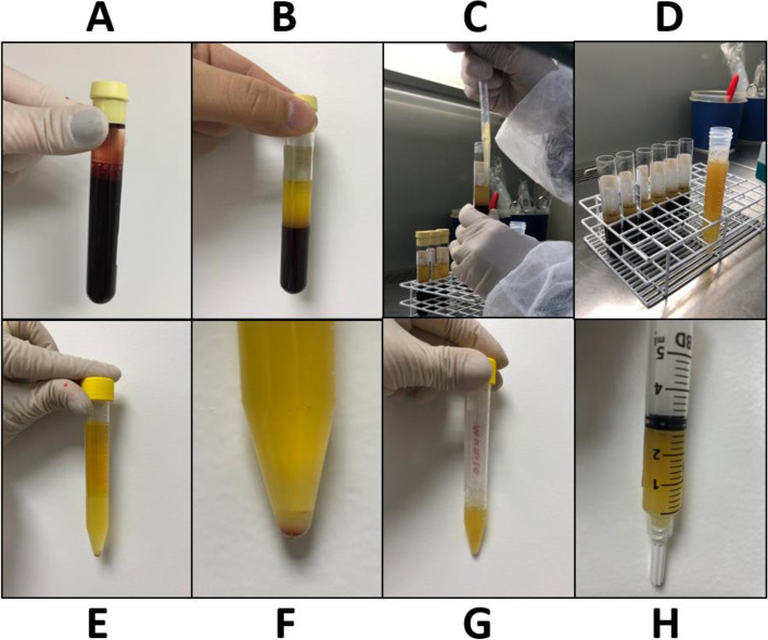
Fig. 1.
Preparation of platelet-rich plasma (PRP) and plasma: A – blood collected; B – after 1st centrifugation, separation of erythrocytes on the bottom, an intermediate thin layer known as buffy coat and a superior layer of plasma; C – separation of plasma from all tubes; D – separated plasma in a centrifuge tube (Falcon®); E – after 2nd centrifugation, separation of a pellet of platelets on the bottom and plasma (without platelets); F – magnification of the pellet of platelets; G – plasma withdrawal (for use as treatment injections) and redilution of the pellet of platelets for obtention of a PRP with approximately 1 x 10⁶ platelets/mm3; H – final PRP for treatment injections

